Sign In
Pricing Creators Affiliate Start Creating
Е

Елена

@elena-4

Сделать своё на Doitong
video
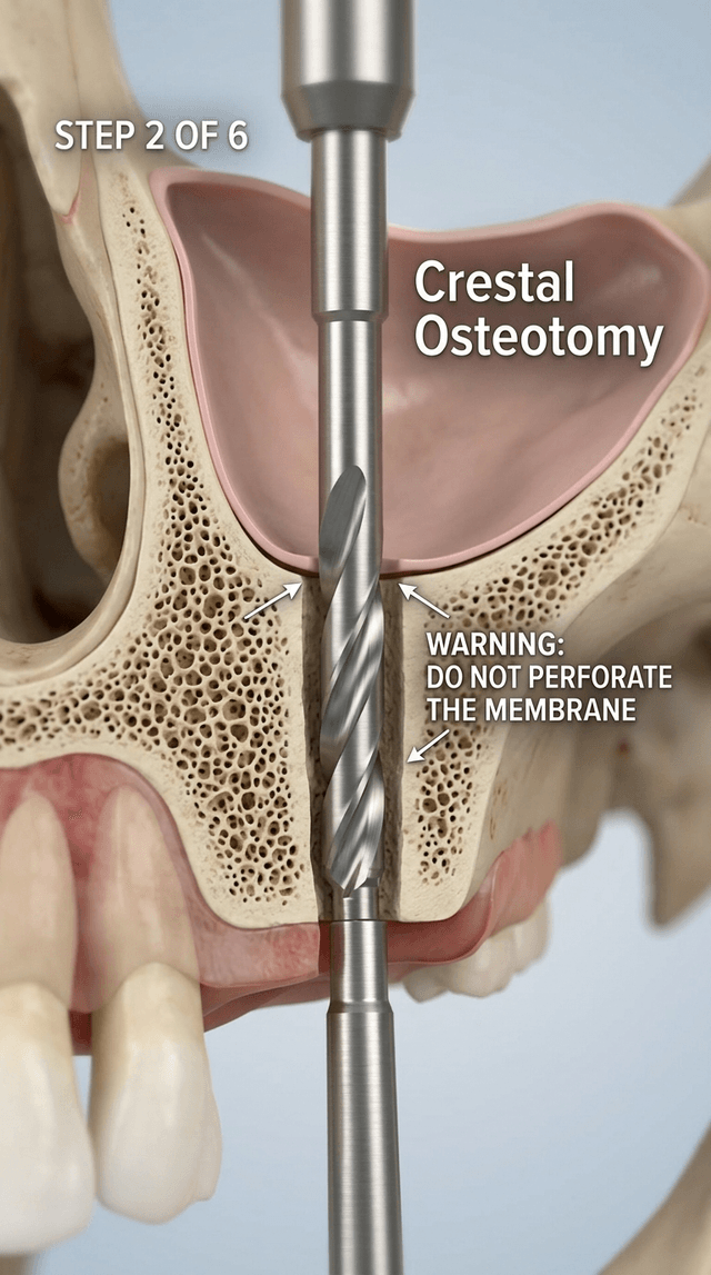
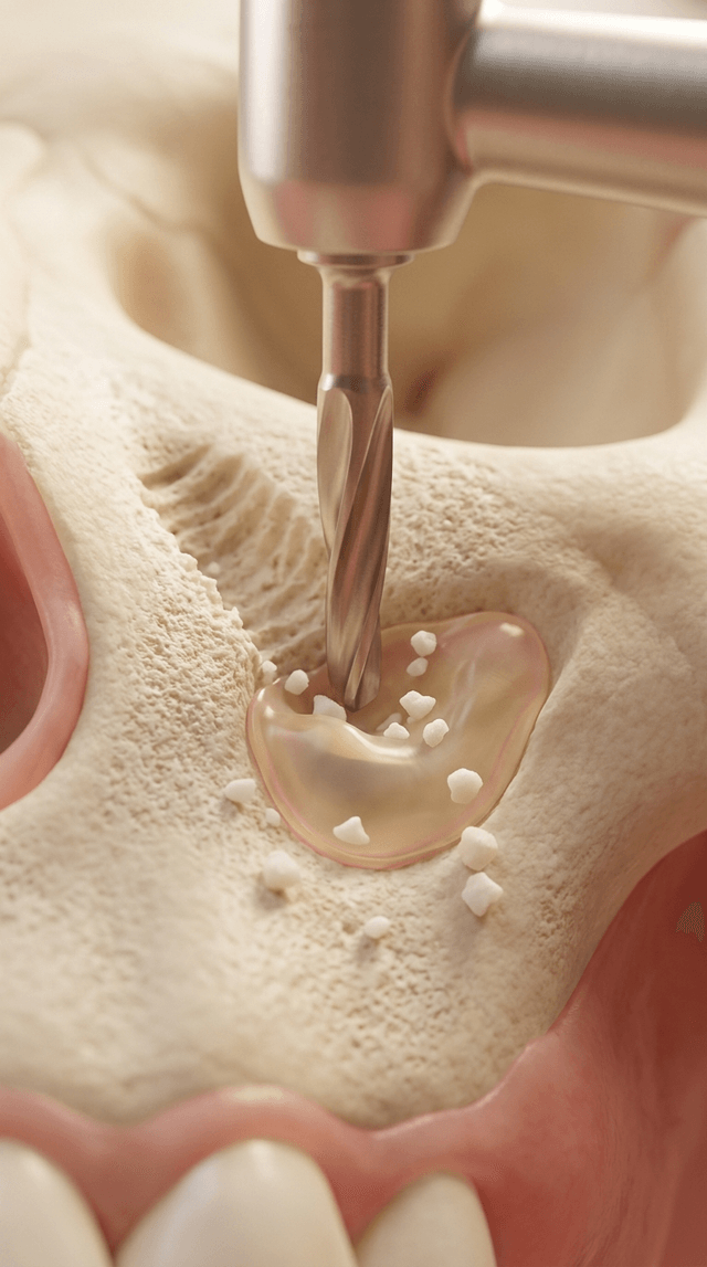
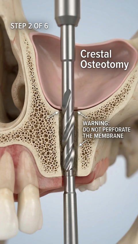
The dental drill bit rotates rapidly and moves upward through the bone crest. The camera remains fixed as the drill stops precisely at th...
image
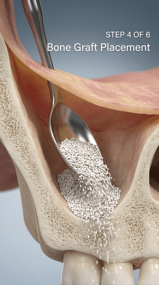
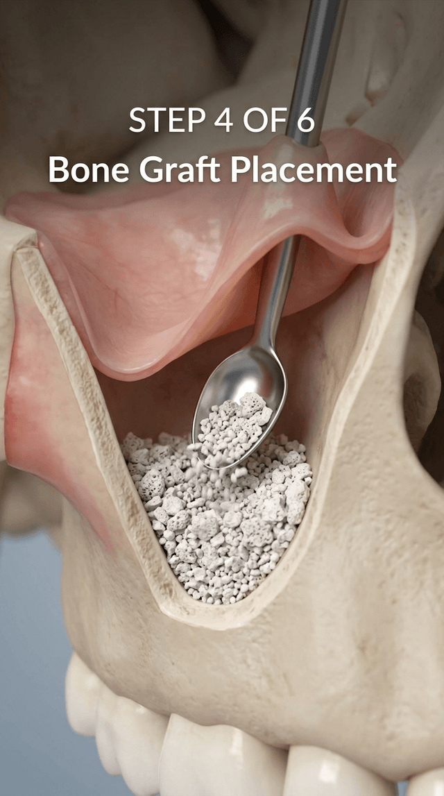
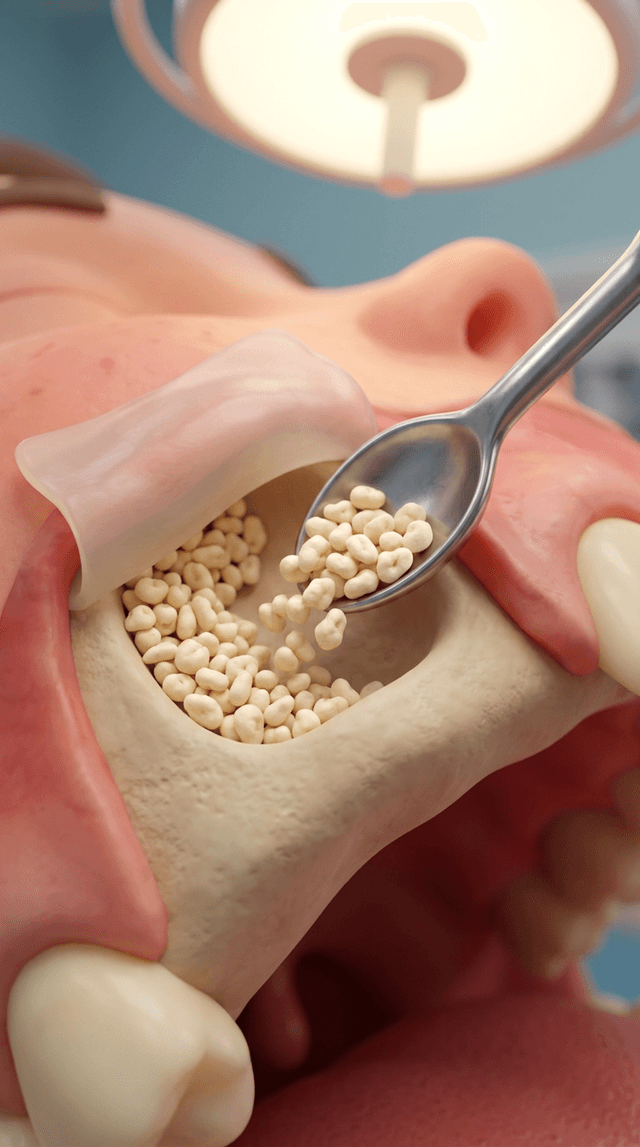
Art style: 3D medical animation. A small metallic spoon-shaped carrier tool is seen inside the created space under the sinus membrane, r...
image
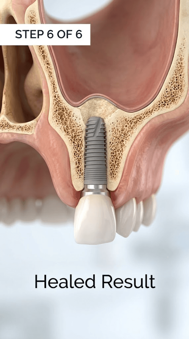
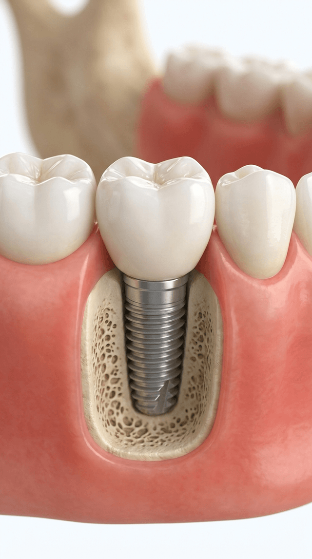
Art style: 3D medical animation. The final result of the procedure, showing the dental implant fully integrated into the bone with a whi...
image
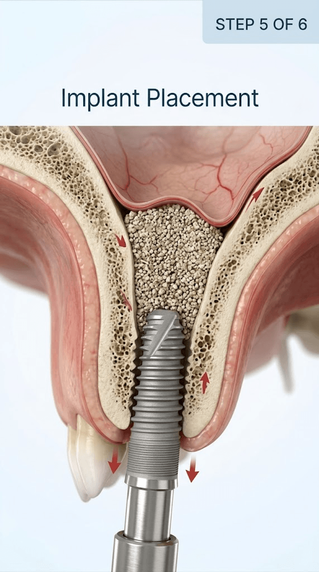
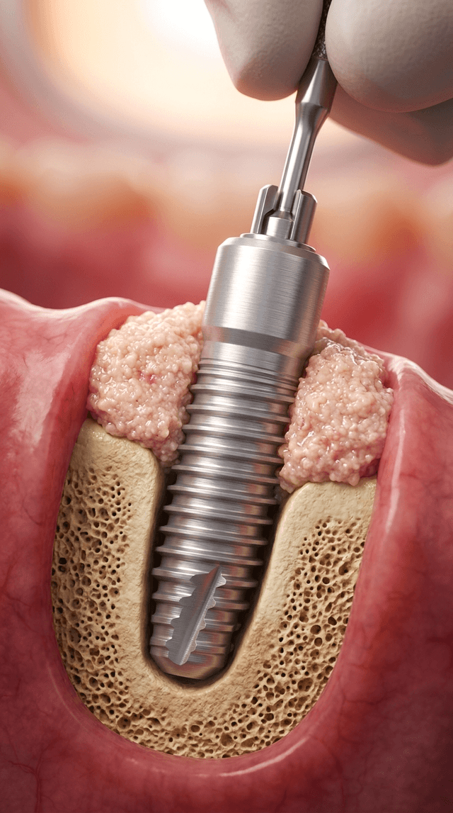
Art style: 3D medical animation. A threaded titanium dental implant is being driven into the bone channel from below, with its tip enter...
image
Art style: 3D medical animation. A closing screen featuring the Instagram logo and the text '@DENTAL_MENTOR' centered on the screen. The...
image
Art style: 3D medical animation. A small metallic spoon-shaped carrier tool is seen inside the created space under the sinus membrane, r...
image
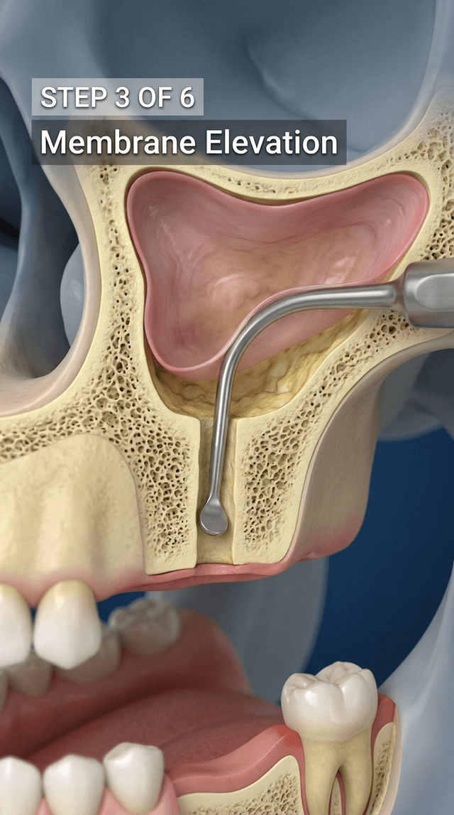
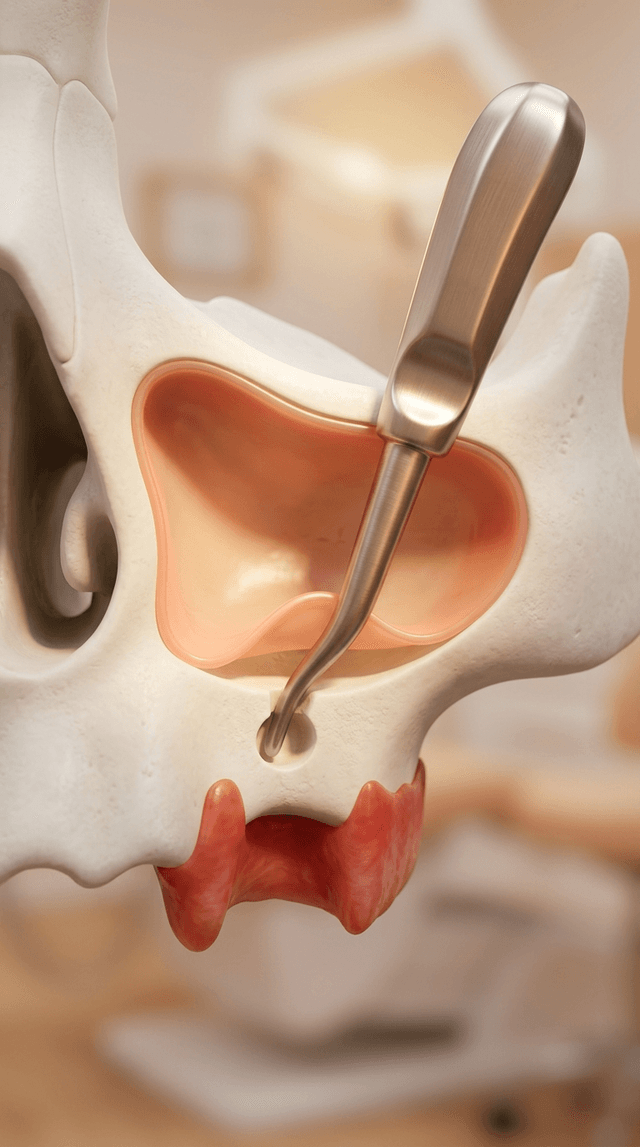
Art style: 3D medical animation. A specialized curved dental elevator tool is inserted through the bone channel, gently lifting the thin...
image
Art style: 3D medical animation. A metallic dental drill bit is positioned inside a vertical channel cut into the jawbone, just below th...
image
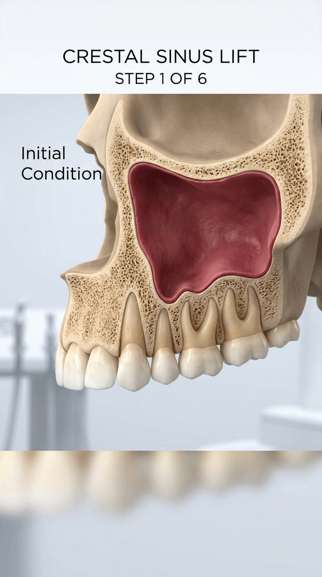
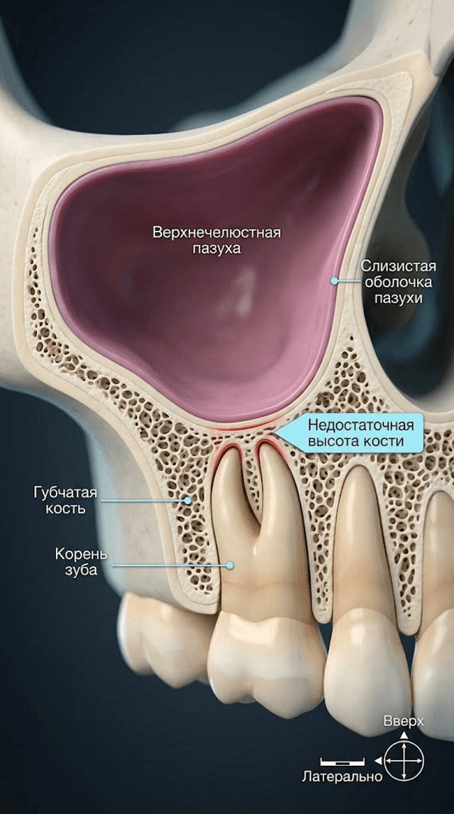
Art style: 3D medical animation. A detailed 3D cross-section of the human upper jaw and maxillary sinus. The bone is porous and tan-colo...
video
Плавная анимация вращения и погружения имплантата в кость до полной установки. Камера немного поворачивается, чтобы показать стабильность...
image
Art style: 3D Pixar. 3D-медицинская анимация. Полностью интегрированный имплантат в зажившей кости. Сверху на имплантат устанавливается ...
image
Art style: 3D Pixar. 3D-медицинская анимация. Титановый зубной имплантат вкручивается в кость и в массу костного трансплантата. Крупный ...
video
Плавная анимация движения сверла вниз, создающего отверстие в кости. Камера следует за кончиком сверла. On-screen dialogue (characters s...
image
Art style: 3D Pixar. 3D-медицинская анимация. Инструмент, похожий на ложку, вносит гранулированный костный материал в пространство под п...
image
Art style: 3D Pixar. 3D-медицинская анимация. Специальный изогнутый стоматологический инструмент (элеватор) осторожно вводится в отверст...
image
Art style: 3D Pixar. 3D-медицинская анимация. Стоматологическое сверло входит в кость, останавливаясь прямо под мембраной Шнайдера. Видн...
video
Статичный кадр с медленным приближением для акцентирования внимания на проблемной области. Текст появляется на экране. On-screen dialogu...
image
Art style: 3D Pixar. 3D-медицинская анимация, поперечное сечение челюсти. Видна верхнечелюстная пазуха, губчатая кость и зуб. Недостаточ...
video
b4beac0eebac4f63a0a83d02a5ae24fb.mp4
image
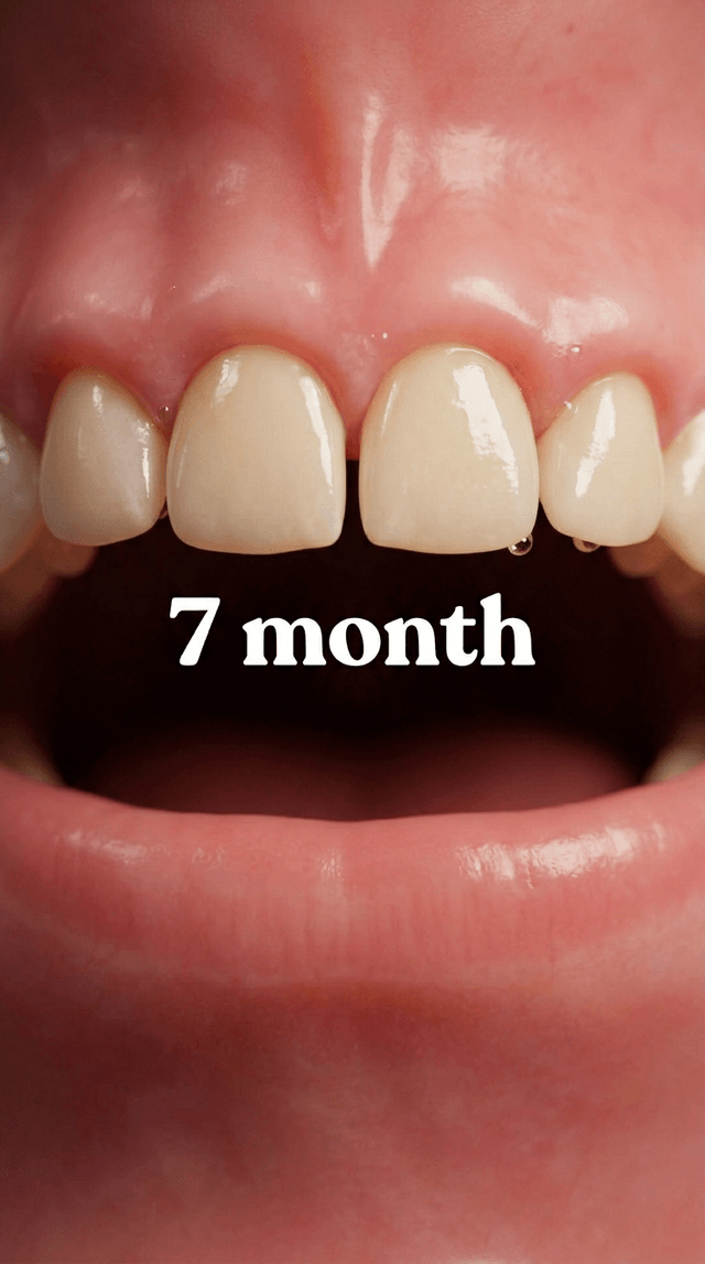
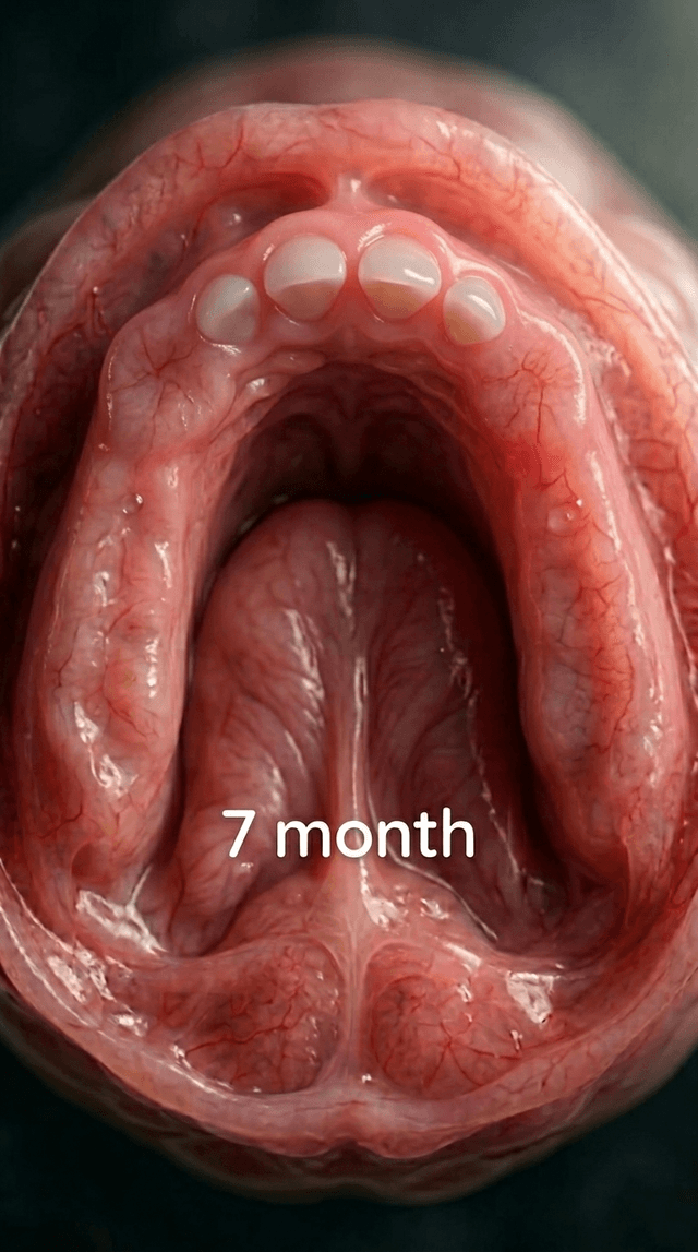
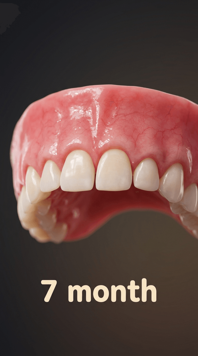
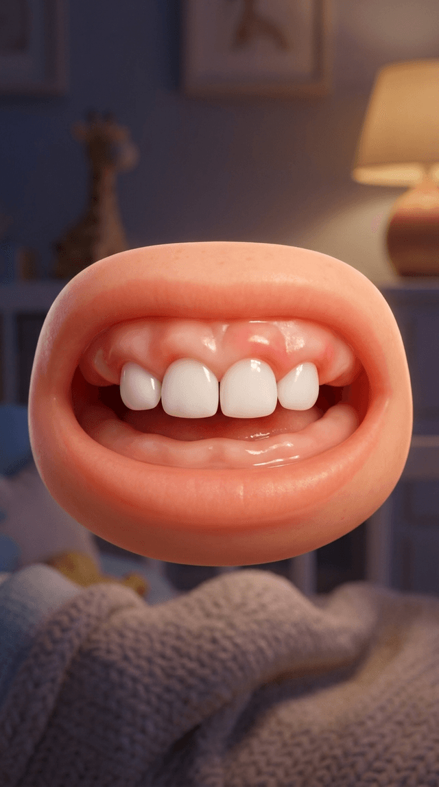
Art style: 3D Pixar. A macro view of the upper dental arch at seven months. Four front teeth, the central and lateral incisors, are now ...
image
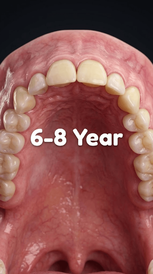
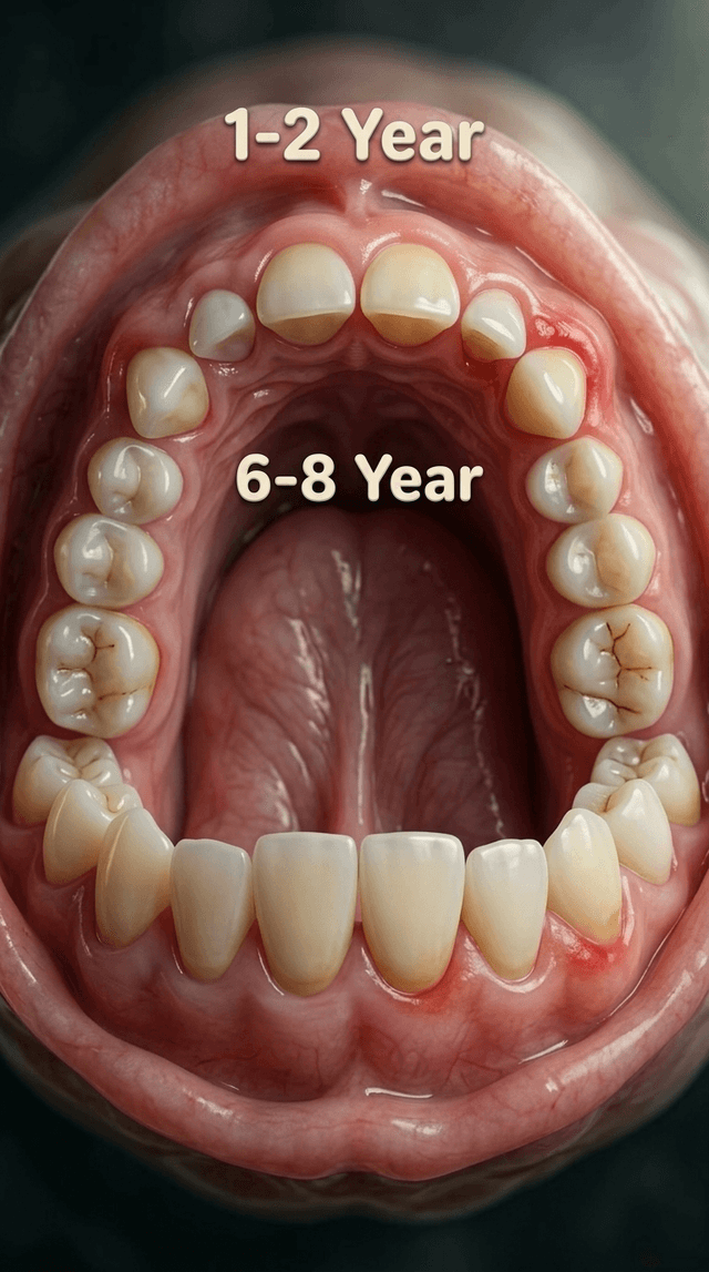
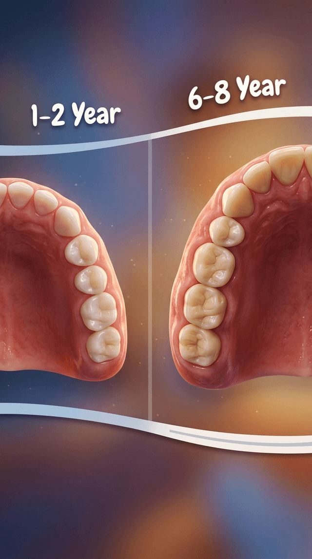
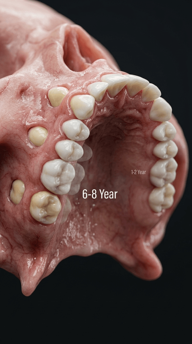
Art style: 3D Pixar. A close-up view of the upper palate at six to eight years. The baby teeth are being replaced by larger, permanent a...
image
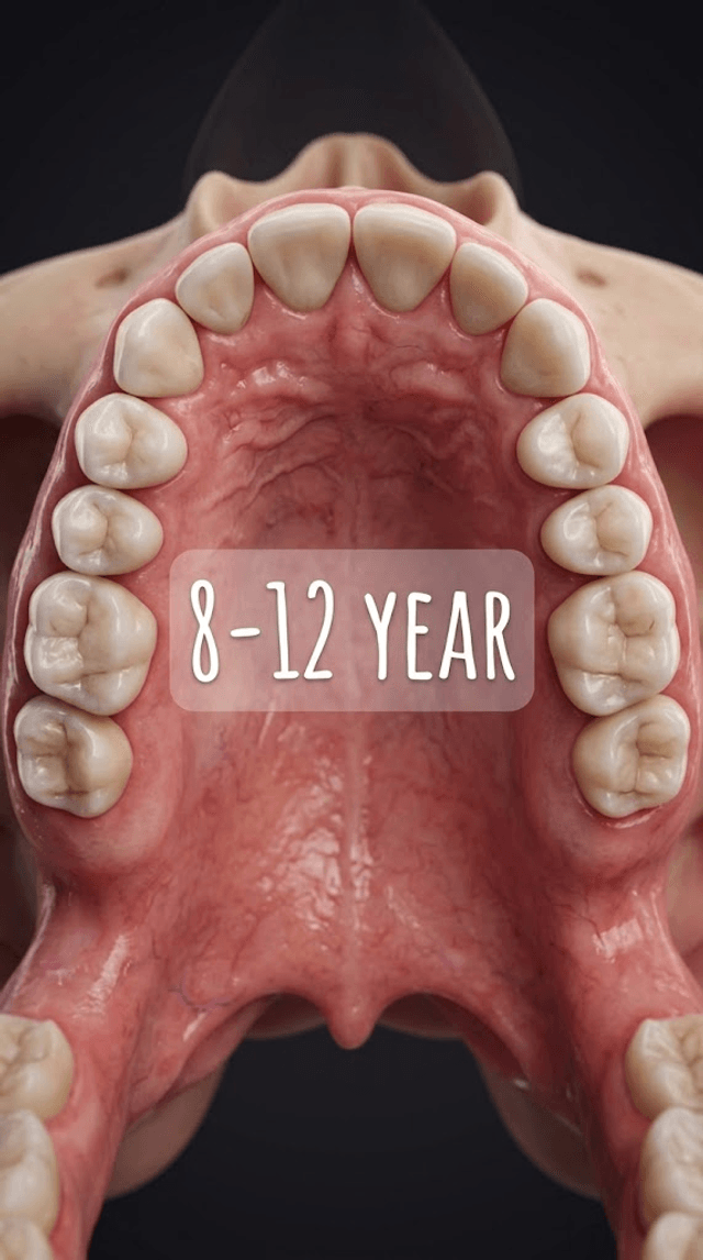
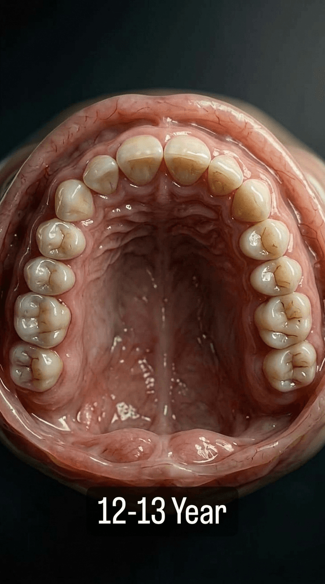
Art style: 3D Pixar. A close-up view of the upper palate at eight to twelve years. Most of the primary teeth have been replaced by perma...
image
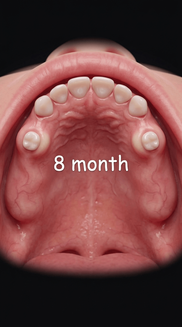
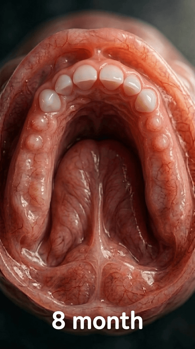
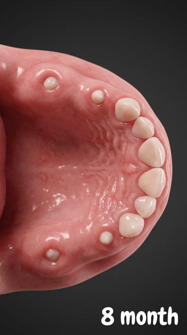
Art style: 3D Pixar. A close-up view of the upper palate at eight months. In addition to the four front teeth, two small white bumps are...
image
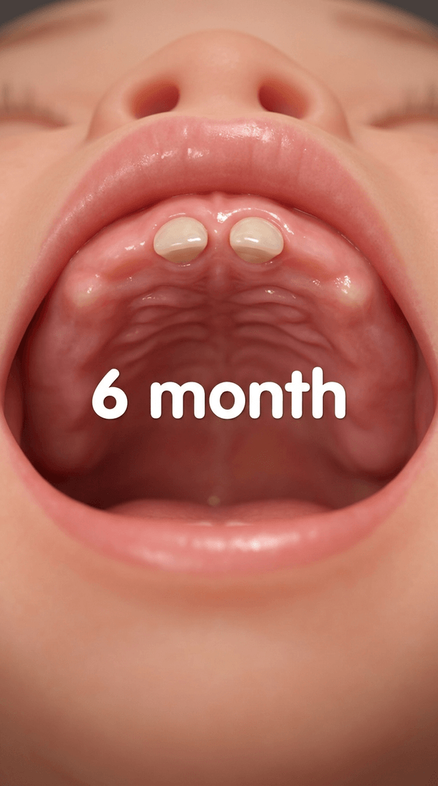
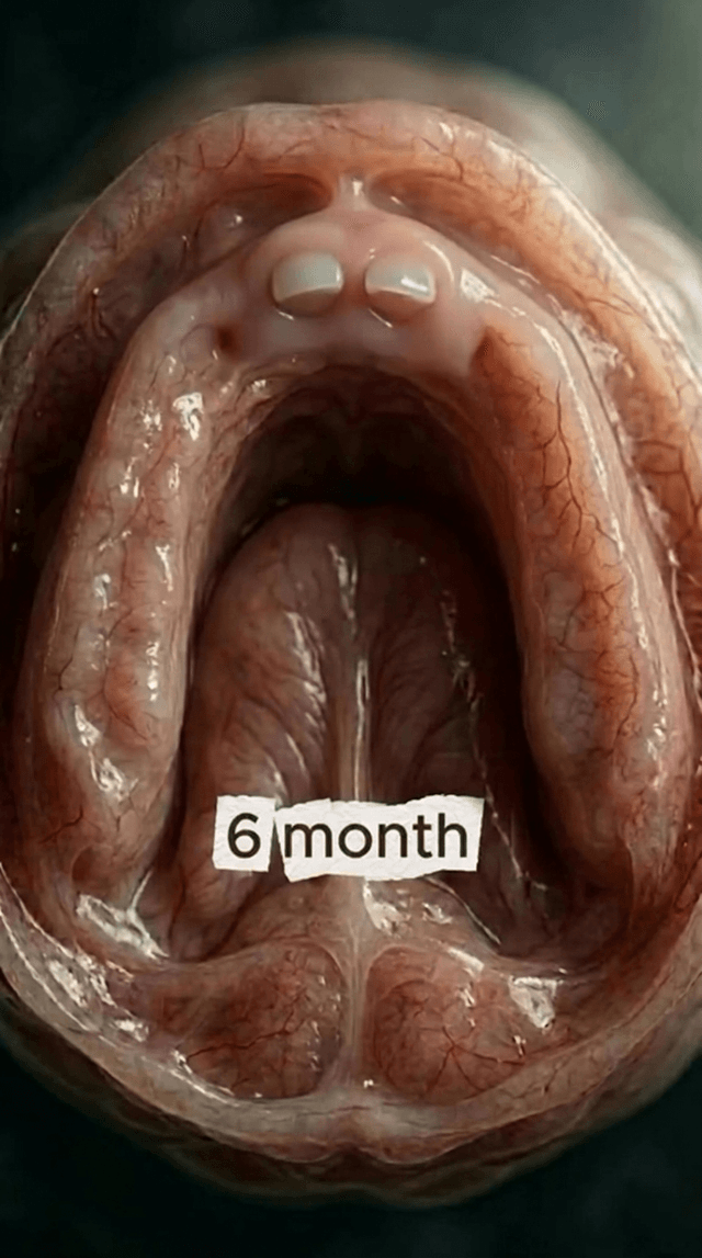
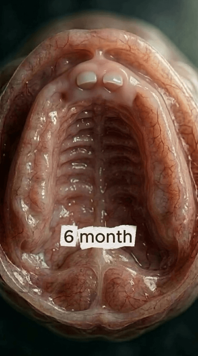
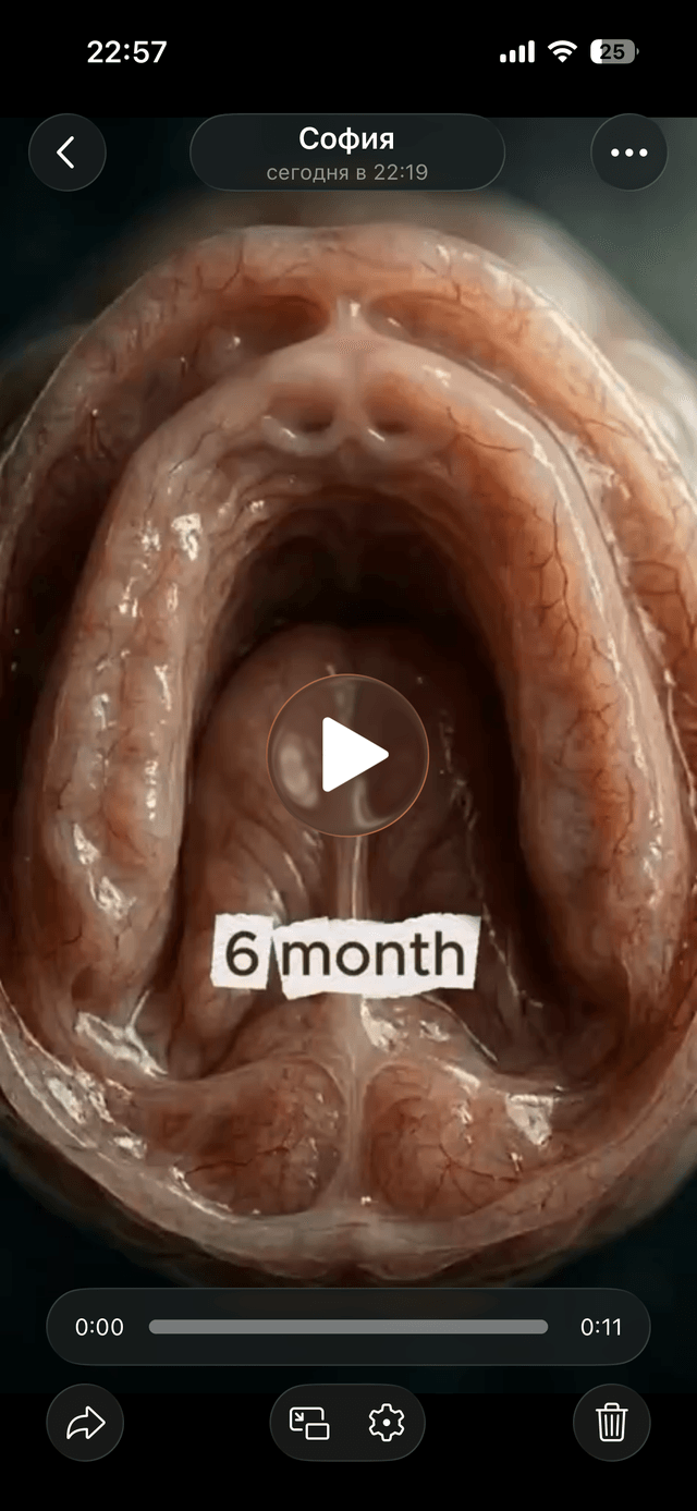
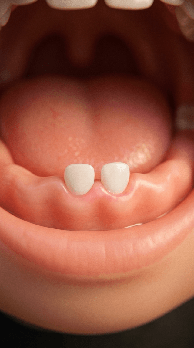
Art style: 3D Pixar. A close-up, bottom-up view of a baby's upper palate. The gums are smooth, healthy pink, and moist. Two small, trans...
image
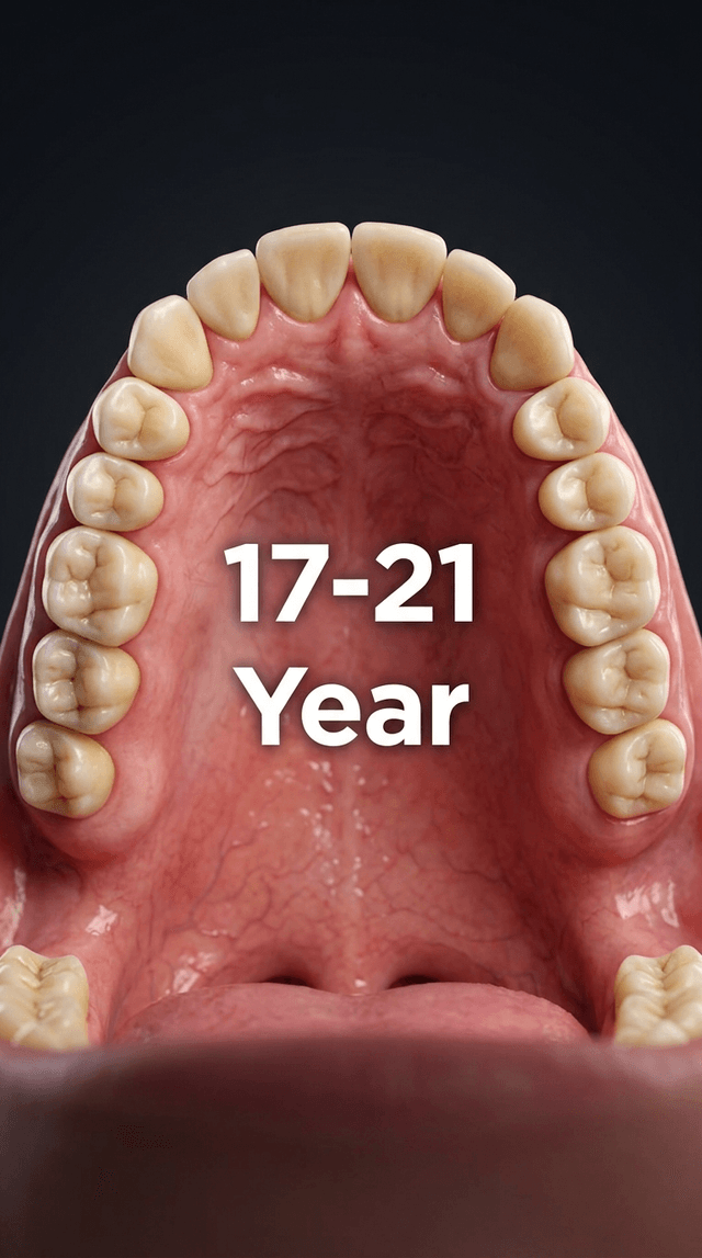
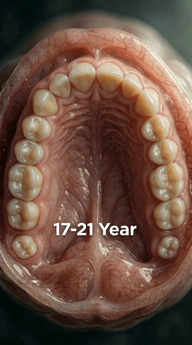
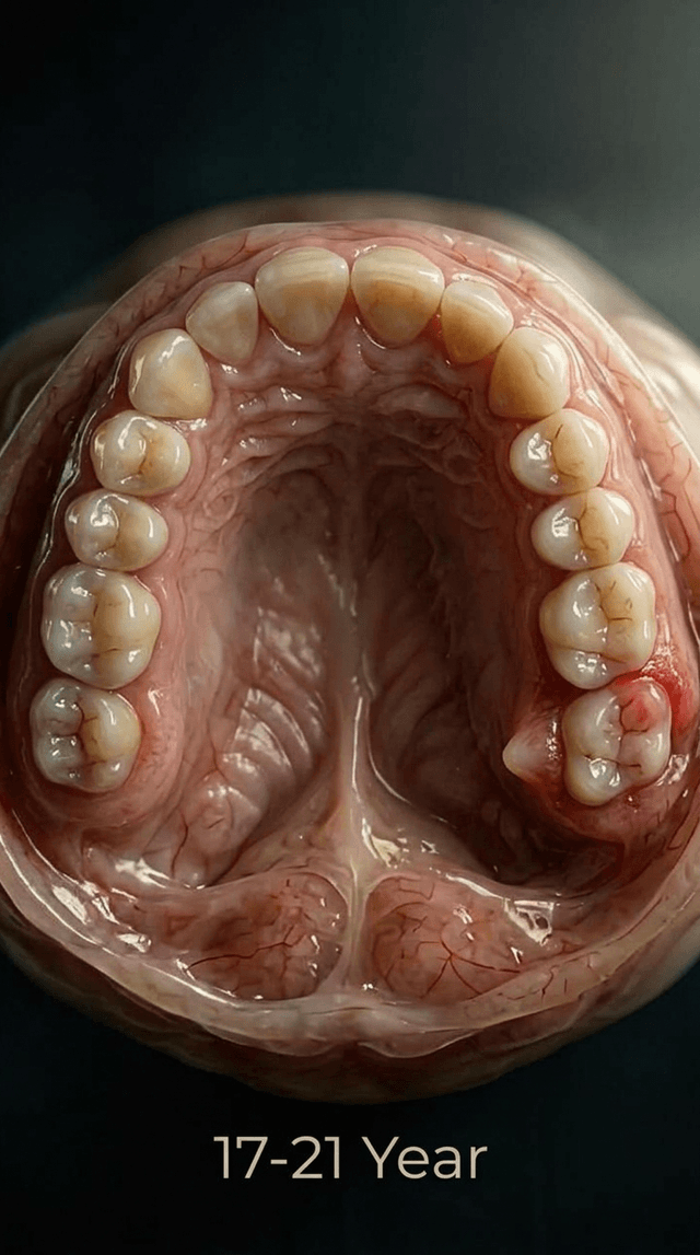
Art style: 3D Pixar. A close-up view of the adult upper palate at seventeen to twenty-one years. The third molars, or wisdom teeth, are ...
image
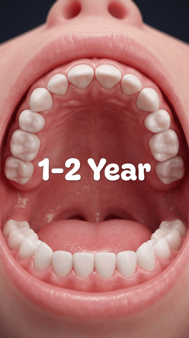
Art style: 3D Pixar. A close-up view of the upper palate at one to two years. A full set of primary baby teeth is visible, including inc...
image
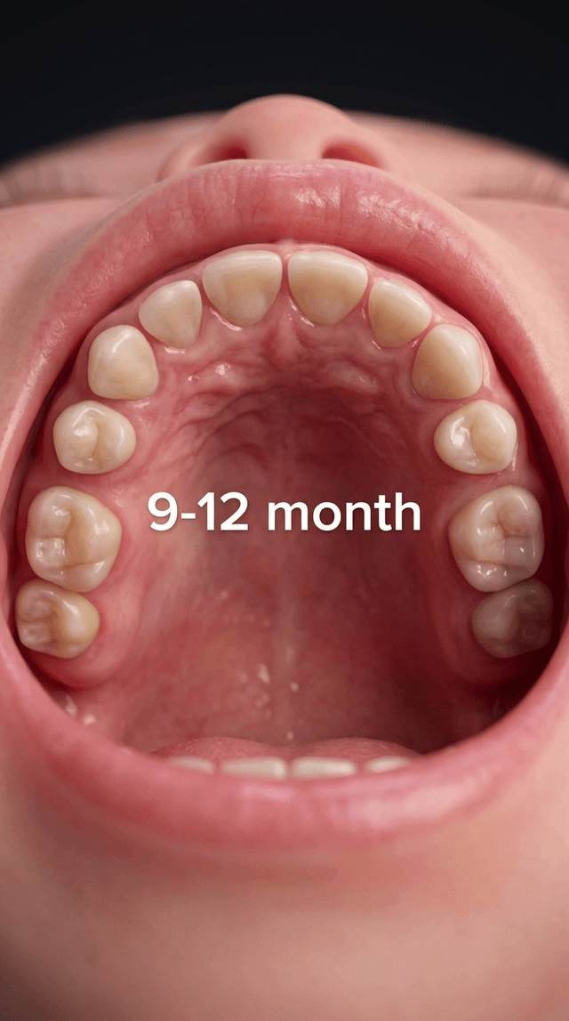
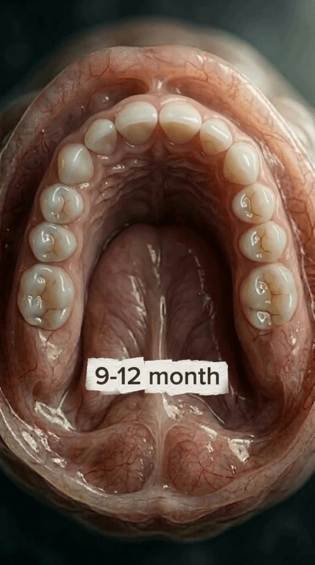
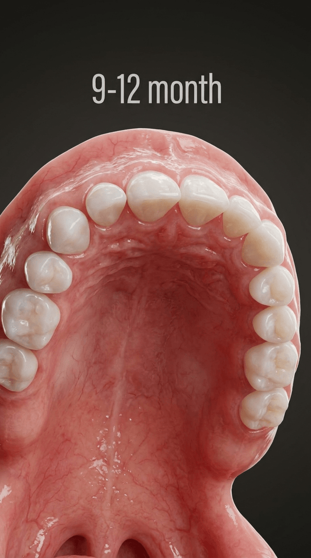
Art style: 3D Pixar. A close-up view of the upper palate between nine and twelve months. More teeth are present, including the lateral i...
image
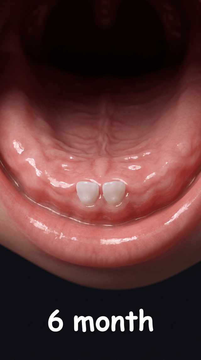
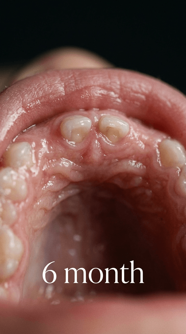
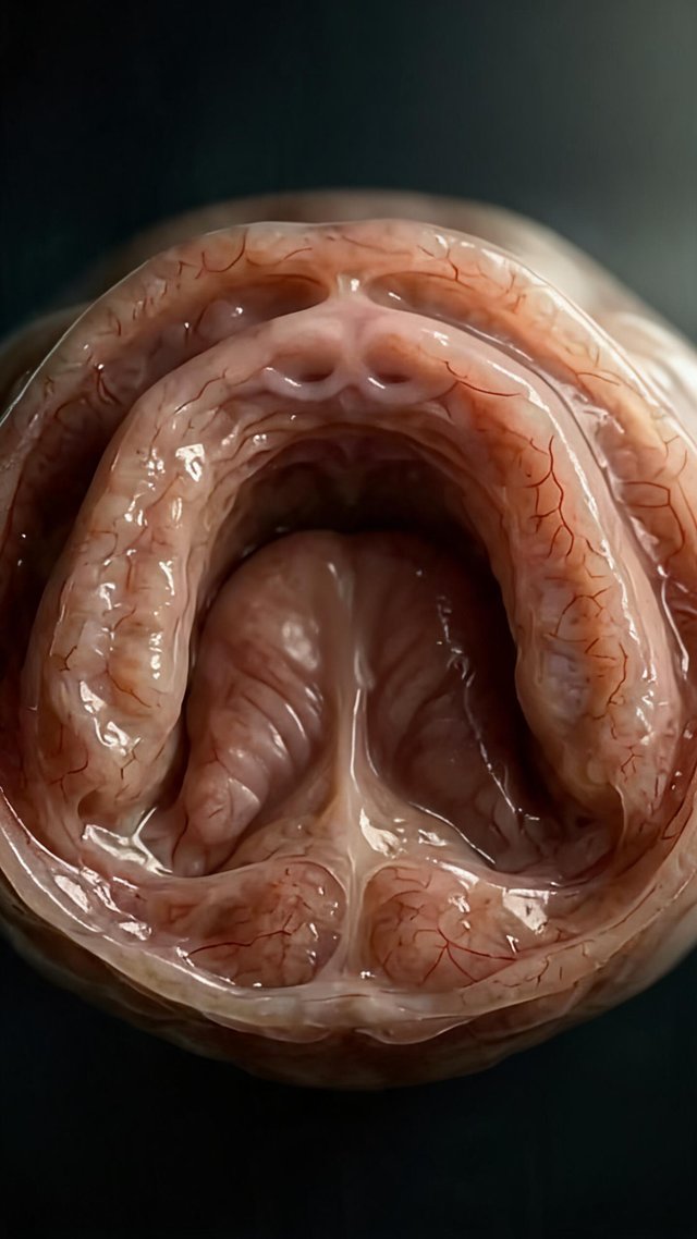
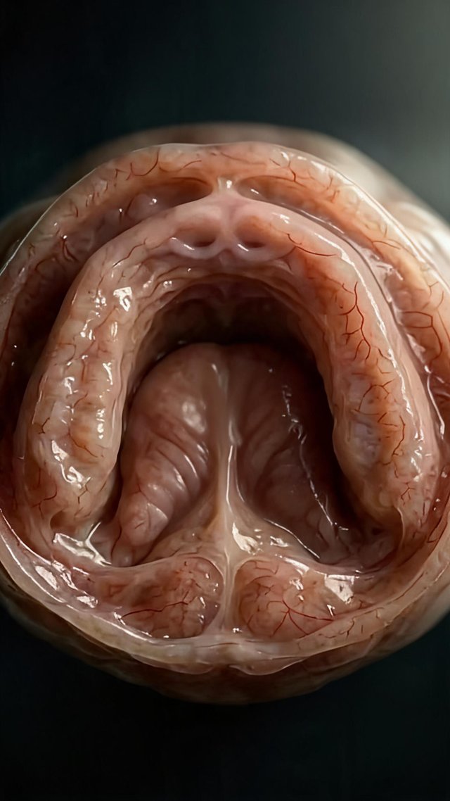
Art style: 3D Pixar. A detailed, anatomical view of a human upper jaw at six months of age. The central focus is the hard palate, which ...
image
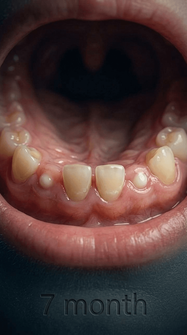
Art style: 3D Pixar. The anatomical view of the upper jaw at seven months. Four teeth are now visible along the front of the dental arch...
image
Art style: 3D Pixar. The upper jaw during the transition from early childhood to the start of permanent dentition. The primary teeth are...
image
Art style: 3D Pixar. A detailed, anatomical view of a human upper jaw at six months of age. The central focus is the hard palate, which ...
image
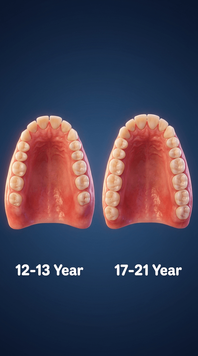
Art style: 3D Pixar. The upper jaw in late adolescence and early adulthood. A full set of permanent teeth is present, including the prem...
image
Art style: 3D Pixar. The upper jaw between nine and twelve months of age. A more complete set of primary teeth is now visible, with the ...
image
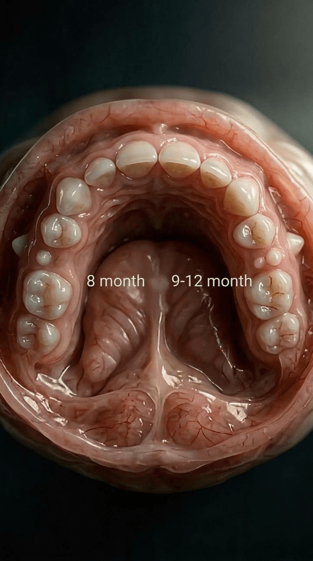
Art style: 3D Pixar. The upper jaw at eight months of age. The four front incisors are fully emerged, and new activity is visible furthe...
image
Art style: 3D Pixar. View of the upper jaw between 9 and 12 months. Most primary teeth are erupting, creating a nearly full set of baby ...
image
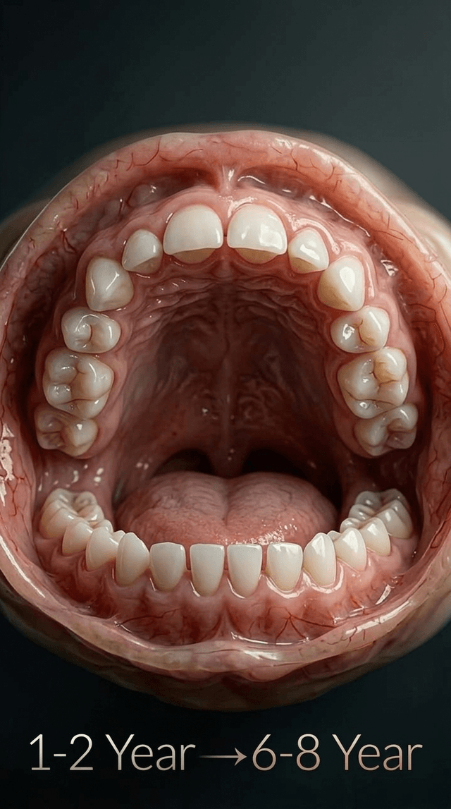
Art style: 3D Pixar. Transitioning view of the upper jaw from 1 year to 8 years. The smaller primary teeth are being replaced by larger,...
image
Art style: 3D Pixar. Anatomical view of the upper jaw at 7 months. Four central and lateral incisors are now clearly visible, protruding...
image
Art style: 3D Pixar. Close-up of an infant's upper jaw at 6 months. Two small, white central incisors are just beginning to erupt throug...
image
Art style: 3D Pixar. Detailed view of the upper jaw at 8 months. The incisors are further developed, and small white points for the firs...
image
Art style: 3D Pixar. Final development of the upper jaw from 8 to 21 years. The full adult dental arch is complete, including the wisdom...
image
Art style: Cinematic Realistic. A view of the upper palate with a full set of primary teeth. The teeth are small, white, and evenly spac...
image
Art style: Cinematic Realistic. A close-up view of an infant's upper palate. The pink, fleshy gums are smooth and moist. Two tiny white ...
image
Art style: Cinematic Realistic. The upper jaw of a seven-month-old infant. The two central incisors have partially erupted through the p...
image
Art style: Cinematic Realistic. The upper jaw showing more advanced tooth eruption. Four front teeth are now visible, and the first mola...
image
Art style: Cinematic Realistic. A fully developed adult upper jaw. The third molars, or wisdom teeth, are erupting at the very back of t...
image
Art style: Cinematic Realistic. A view of the upper palate with a full set of primary teeth. The teeth are small, white, and evenly spac...
image
Art style: Cinematic Realistic. The developing adult upper jaw. The permanent incisors are fully erupted, and the premolars and second m...
image
IMG_0427.jpeg
video
977835774244066685.mp4
image
First frame
image
IMG_0426.jpeg
image
IMG_0426.jpeg
video
977835774244066685.mp4
video
977835774244066685.mp4
video
977835774244066685.mp4
video
977835774244066685.mp4
image
IMG_0403.png
image
Art style: 3D Pixar. Та же 3D-модель челюсти. Теперь видны четыре верхних резца. Они маленькие, аккуратные и идеально белые. Десны вокру...
image
Art style: 3D Pixar. Стилизованная 3D-модель детской челюсти, вид изнутри. Десны нежно-розовые, гладкие. Из десны едва-едва прорезаются ...
video
977835774244066685.mp4